Syllabus (Fourth Edition, 2023)
Topics
i. Explain the ionic basis of spontaneous electrical activity of cardiac muscle cells.
ii. Describe the normal processes of cardiac excitation and electrical activity.
iii. Correlate the mechanical events of the cardiac cycle with the physical, electrical, and ionic events
Topics not covered in previous SAQs
.
Learning Objectives for the First Part Examination in Intensive Care Medicine
- This will ensure that trainees, tutors, and examiners can work from a common base.
- All examination questions are based around this Syllabus.
- These learning objectives are designed to outline the minimum level of understanding required for each topic.
- The accompanying texts are recommended on the basis that the material contained within them provides sufficient information for trainees to meet the learning objectives.
- Trainees are strongly encouraged to explore the existing and evolving body of knowledge of the Basic Sciences as they apply to Intensive Care Medicine by reading widely.
- For all sections of the syllabus an understanding of normal physiology and physiology at extremes of age, obesity, pregnancy (including foetal) and disease (particularly critical illness) is expected.
- Similarly, for pharmacology, trainees are expected to understand a drug’s pharmacology in these contexts.
- An understanding of potential toxicity and relevant antidotes is also expected.
Definitions
Throughout the document specific wording has been used under the required abilities to indicate the level of knowledge and understanding expected and a glossary of these terms is provided.
Definitions
| Calculate | Work out or estimate using mathematical principles. |
| Classify | Divide into categories; organise, arrange. |
| Compare and contrast | Examine similarities and differences. |
| Define | Give the precise meaning. |
| Describe | Give a detailed account of. |
| Explain | Make plain. |
| Interpret | Explain the meaning or significance. |
| Outline | Provide a summary of the important points. |
| Relate | Show a connection between. |
| Understand | Appreciate the details of; comprehend. |
SAQs
i. Explain the ionic basis of spontaneous electrical activity of cardiac muscle cells.
ii. Describe the normal processes of cardiac excitation and electrical activity.
2022B 05
Outline the structure of fast cardiac sodium channels and describe in detail how they work.
CICMWrecks Answer
Sodium channels
Classified into:
- Voltage-gated sodium channels
- In exitable cells such as myocytes, neurons, certain glia
- Very selective to Na+
- Responsible for rising phase of action potentials
- Triggered by change in membrane potential (voltage)
- Ligand-gated sodium channels
- NMJ – Nicotinic receptors (ACH – ligand)
- Permeable to Na+ and K+
- Triggered by binding of ligands to channel
- Leak sodium channel
- Ungated, always open
- Contributes to depolarization from K+ potential
- Increasing permeability lowers RMP, brining it closer to trigger of an action potential
Voltage-gated Sodium Channel
- Family consisting of Nav 1.1-1.9 encoded by genes SCN1A to SCN9A respectively
- Human Cardiac Sodium channel Nav 1.5 encoded by SCN5A
- responsible for the generation of the rapid upstroke of the myocardial action potential
- determines impulse conduction velocity in cardiac tissue (affects action potential upstroke velocity — extent of intercellular communication via gap junctions)
- Mutations in the gene encoding this channel (SCN5A) linked to three forms of primary electrical disease (channelopathies)
- the long QT syndrome (LQTS)
- the Brugada syndrome (BS) and
- cardiac conduction defects such as broad QRS, LBBB/RBBB, prolonged PR intervals, and other channelopathies like familial AF and idiopathic VF.
Structure
composed of an α subunit (forms ion conduction pore) and one or more auxiliary β subunits (several functions incl. modulation of channel gating)
α subunit
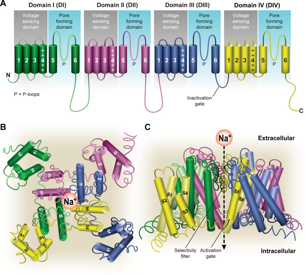
- α subunit is the largest subunit and contains both the pore region of the channel and the voltage sensor
- forms core of the channel and is functional on its own
- indepently able to form a pore in the cell membrane that conducts Na+ in a voltage-dependent way
- When accessory proteins assemble with α subunits, the resulting complex can display altered voltage dependence and cellular localization.
- Principal α-subunit composed of four homologous domains (DI–DIV), each containing six transmembrane segments (S1–S6). The four domains are attached to one another by cytoplasmic linker sequences.
- Highly conserved S4 segment acts as the channel’s voltage sensor
- When stimulated by a change in transmembrane voltage, this segment moves toward the extracellular side of the cell membrane, forming the central pore cavity and allowing the channel to become permeable to ions
- Central Pore Cavity
- Conduct ions
- External portion(i.e., more extracellular): formed by the “P-loops” (the region between S5 and S6) of the four domains. This region is the most narrow part of the pore and is responsible for its ion selectivity (Selectivity filter). Keep out anions and large cations (K+)
- Inner portion (i.e., more cytoplasmic): is the pore gate and is formed by the combined S5 and S6 segments of the four domains.
- Also features lateral tunnels or fenestrations: that run perpendicular to the pore axis. Connect the central cavity to the membrane. Proposed to be important for drug accessibility.
Gating
- have two gates
- Activating gate (m) (voltage-dependent): Opening of the activating gate allows the influx of sodium and cell depolarization.
- Inactivation gate (h) (time-dependent): closing of the inactivation gate will stop the flow of sodium regardless of the status of the activation gate
- Highly conserved S4 segment acts as the channel’s voltage sensor
- When stimulated by a change in transmembrane voltage, this segment moves toward the extracellular side of the cell membrane, forming the central pore cavity and allowing the channel to become permeable to ions
- The cytoplasmic linker between domains III and IV of the α subunit is responsible for inactivation of the Na channel.
- After the channel opens, this segment of the protein is drawn to the inner surface of the channel like a “hinged lid,” – physically wedges the pore gate shut – blocks Na+ ions from passing through the pore
- 3 main conformational states: open, closed, inactivated
- Ion permeable only in open state
- Open: m gates open rapidly
- Closed(resting): m closed, h open
- Inactive:
- Transitions:
- Activation/Deactivation (Closed ⇌ Open)
- Inactivation/Reactivation (Open ⇌ Inactivated)
- Recovery from inactivation/closed-state inactivation (Inactivated ⇌ Closed)
Ionic Events
| Action Potential | Membrane Potential | Target Potential | Gates | NA+ / RP | Gate’s Target State | Phase of Cardiac AP |
|---|---|---|---|---|---|---|
| Resting | −90 mV | −70 mV | m closed | Ready to go | Deactivated → Activated | Phase 4 |
| Rising | −70 mV | 0 mV | m open rapidly (0.1-0.2sec) | Na flow in | Activated | Phase 0 |
| Rising | 0 mV | +20 mV | h starts closing | Na flow slows | Activated → Inactivated | Phase 0 |
| Falling | +20 mV | 0 mV | h closed (takes 10msec) | Na+ channel refractory period starts | Inactivated | Phase 1 |
| Falling | 0 mV | −90 mV | h remains closed | RP ongoing | Inactivated | Phase 2,3 |
| Undershot | −90 mV | −95 mV | h open, m closes | RP ends | Inactivated → Deactivated | Phase 4 |
| Rebounding | −95 mV | −90 mV | m closed | ready to go | Deactivated | Phase 4 |
β subunit
- Four different protein isoforms
- each about one-tenth of the mass of the α subunit
- anchored in the cell membrane through one transmembrane segment
- Extracellular domains are large and have structures similar to those of immunoglobulins.
- Regulate the expression and subcellular targeting of Na channels, and modulate the function (gating) of the α subunit
Examiner Comments
2022B 05: 18% of candidates passed this question.
The expectation for this question was an appropriate description of the fast sodium channel structure included outlining its single alpha and 2 beta sub-units, activation (m) and inactivation (h) gates and sodium selectivity. It was expected that candidates would comment on cycling between the 3 states (resting, open and inactive), corresponding conformational changes to the fast sodium channel, triggers for these changes (voltage or time) and ionic events, including reference to and description of absolute and relative refractory periods. Better candidates were able to relate events to a diagram of a fast action potential, simply drawing a diagram without referencing events at the sodium channel scored few marks. No marks were awarded for description of drug effects on the fast sodium channel. The question specifically relates to fast Na channels, so descriptions of other channels (e.g. ligand gated sodium channels) did not score marks. Details of the subsequent contraction of cardiac muscle following an action potential was also not part of the question and did not score marks.
2024A 12
(a) Describe the action potential of the cardiac pacemaker cells including the ionic events (60%).
(b) Explain how excitation then propagates through the conducting pathway of the heart, including mechanisms to prevent abnormal conduction (40%).
2008A 19
Outline normal impulse generation and conduction in the heart.
Describe the features present in a normal heart that prevent generation and conduction of arrhythmias.
CICMWrecks Answer
Normal Impulse Generation
Occurs in the sinoatrial node
- Innervated by (predominantly right) vagal nerve and sympathetic nerves
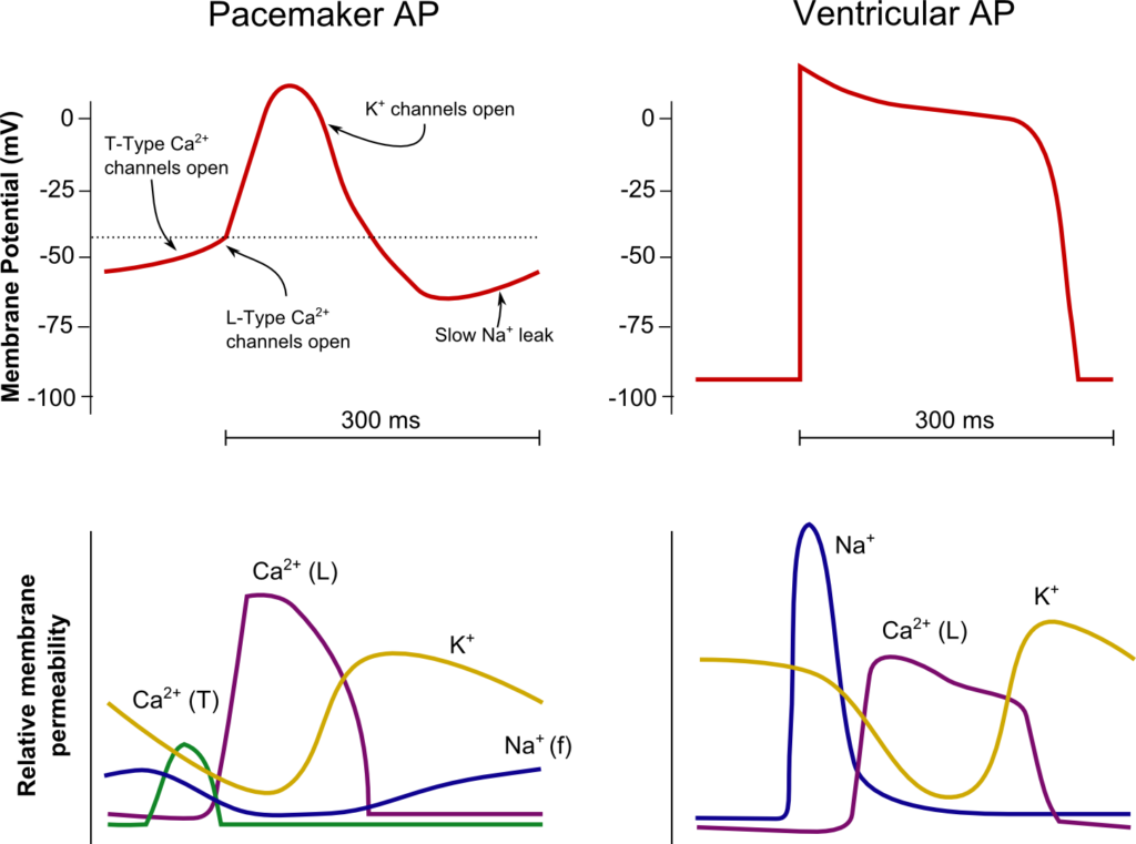
- Upslope of phase 4 due to “Funny” Na current (inward flow of Na)
- Parasympathetic (vagal) stimulation causes decrease (more negative) in the minima and shallower upward gradient of phase 4, delaying depolarization
- Sympathetic stimulation causes an increase (more positive) in the minima and steeper upward gradient of phase 4, hastening depolarization
- Action potential generated when threshold potential reached (~-50-55mV) → Opening of voltage gated Ca channels → Ca influx
- Cell begins repolarization when Ca channels close and K channels open à K efflux
Normal Conduction
- SA node (5cm/sec) → Internodal fibres and Bachmann’s plexus (100cm/sec) → AV node (5cm/sec) → Bundle of His (100cm/sec) → Purkinje fibres (400cm/sec)
- Occurs due to fast wave action potentials (slow wave in AV node)
Ventricular AP:
- Phase 0 (Fast upstroke)
- Occurs due to opening of voltage gated Na channels (Na influx)
- Overshoot to +20mV
- Occurs due to opening of voltage gated Na channels (Na influx)
- Phase 1 (Early repolarization)
- Inactivation of Na channels and opening of K channels (K efflux) → Start of absolute refractory period
- Phase 2
- Opening of voltage gated Ca channels (Ca influx) balances K efflux
- Phase 3
- Closure of Ca channels → Repolarization
- Na channels transition to closed state (absolute refractory period transitions to relative refractory period)
- Phase 4
- Resting membrane potential (-80mV~-90mV) maintained by Na/K ATPase
Features which Prevent Arrhythmias / Conduction Abnormalities
Preventing the generation of arrhythmias
- SA nodal rate has faster intrinsic firing rate than other areas (~30bpm) causing
override inhibition of other pacemakers
Preventing the conduction of arrhythmias
- AV node prevents the conduction of impulses >220bpm
- Normal myocyte action potential has absolute (where no stimulus can initiate a new
action potential) and relative (where a greater than normal stimulus is required for initiation of a new action potential) refractory periods - Fibrous cardiac skeleton prevents conduction of action potentials other than through
the specialized conduction pathways
Sakurai 2016
Examiner Comments
2024A 12: 69% of candidates passed this question.
(a) A detailed description of the ionic events of the phases of the sino-atrial node AP was expected. A diagram of the sino-atrial node potential which included the phases and labelled x and y axis was helpful in this description. Some comparison with other cardiac action potentials added depth to answers, as did a brief description of the influences on the action potential such as the autonomic nervous system.
(b) This section required a detailed explanation of impulse propagation both between cardiac myocytes and across the myocardium. Safety mechanisms to be mentioned included anatomical insulation, refractory periods and rate.
2008A 19: 1 candidate (33%) passed this question.
This question required description of the SA node, its primary role and generation of the pacemaker potential and the influence of the autonomic nervous system. A diagram of the conducting pathways, highlighting specialized tissues with fast or slow conduction velocities would have been appropriate. The importance of the AV node in preventing retrograde conduction and high rates conducted to the ventricles (>220 / min) was often neglected in answers. A discussion of the Purkinje Fibres with particular reference to the absolute and relative refractory periods was essential.
Additional marks were awarded for mention of the atrial internodal pathways, conduction within the ventricles from the endocardial to epicardial surfaces and the significance of the compensatory pause in response to ectopic beats.
Syllabus C1b 2.a, b;
Reference: Cardiovascular Physiology, “Electrical Activity of the Heart” (Chapter 2), Berne and Levy.
2020B 01 – 2017B 21 – 2013A 19
Describe and compare the action potentials from cardiac ventricular muscle and the sinoatrial node
2016B 11
Compare the action potentials of a sino-atrial node cell and a myocardial cell.
CICMWrecks Answer

SA Node
Ventricle
Resting membrane potential
- No set RMP, however approx. -60mV
- βadrenergic stimulation causes a less negative RMP and increased slope of the initial upstroke
- muscarinic stimulation causes a more negative RMP and a decreased slope of the initial upstroke
Resting membrane potential
- Approx -90mV
Threshold potential
- Approx 40mV
Threshold potential
- Approx. -50mV
Phase 0
- Funny Na+ current allow leakage of Na+ into the the cell slowly increasing membrane potential to the threshold potential
T and L-type Ca2+ channels (voltage gated) open when threshold potential reached
- Ca2+ influx
- Depolarization
- Shallow upstroke compared to ventricular myocyte
Phase 0
- Rapid depolarization
- Opening of fast Na+ channels
- Influx of Na+
- Overshoot to +20mV
Phase 1
- Early repolarization
- Closure of fast Na+ channels and opening of K+ channels (transient outward, inward rectifier)
- Efflux of K+
Phase 2
- Plateau
- Opening of intially T-type Ca2+ channels and subsequently L-type Ca2+ channels
- Ca2+ influx balances K+ efflux
- Na+ channels in closed state à absolute refractory period
- No action potential can be generated in this state
Phase 3
K+ channels open and Ca2+ channels slowly close
- K+ efflux
- Repolarization
- No plateau phase
Phase 3
- Repolarization
- Closure of Ca2+ channels while K+ channels remain open (delayed rectifier)
- K+ efflux
- Returns potential towards RMP
- Na+ channels transition to inactive state à relative refractory period
- Another action potential can be generated with greater adequate stimulus
Phase 4
- Na+/K+ ATPase and Na+/Ca2+ ATPase maintain ionic gradients
Phase 4
- RMP
- Na+/K+ ATPase and Na+/Ca2+ ATPase maintain ionic gradients
Sakurai 2016
Examiner Comments
2020B 01: 72% of candidates passed this question.
This question details an aspect of cardiac physiology which is well described in multiple texts. Comprehensive answers included both a detailed description of each action potential and a comparison highlighting and explaining any pertinent differences. The question lends itself to well-drawn, appropriately labelled diagrams and further explanations expressed in a tabular form. Better answers included a comparison table with points of comparison such as the relevant RMP, threshold value, overshoot value, duration, conduction velocity, automaticity, ion movements for each phase (including the direction of movement) providing a useful structure to the table. Incorrect numbering of the phases (0 – 4) and incorrect values for essential information (such as resting membrane potential) detracted from some responses.
2017B 21: 95% of candidates passed this question.
This topic was well understood and answered by most candidates. Some candidates had a good knowledge base but missed out on potential marks by failing to compare and contrast. A diagram outlining the various phases was a useful way to approach the question.
2016B 11: 75% of candidates passed this question.
A good answer included a well labelled sketch with a description of ion channels and relative directional flow. When using sketches in an answer they should be correctly labelled, and when used as a comparison with another sketch, the differences should be clear e.g. shape, duration and voltage difference.
2013A 19:
A fundamental aspect of cardiac physiology, that overall was well answered. The majority of candidates used figures to good effect. Candidates are reminded that all figures must be correctly labelled (e.g. X and Y axis, phases of action potential, etc.). Common omissions were those that reflected an adequate depth of knowledge (e.g. some of the current flows).
iii. Correlate the mechanical events of the cardiac cycle with the physical, electrical, and ionic events
2024B 13
(a) Define ventricular diastole, including its usual duration (15% of marks).
(b) Describe the cardiovascular events that occur during this period (85% of marks). (ionic and cellular events not required).
2018A 12 – 2011A 21
Briefly describe the cardiovascular events that occur during ventricular diastole.
CICMWrecks Answer
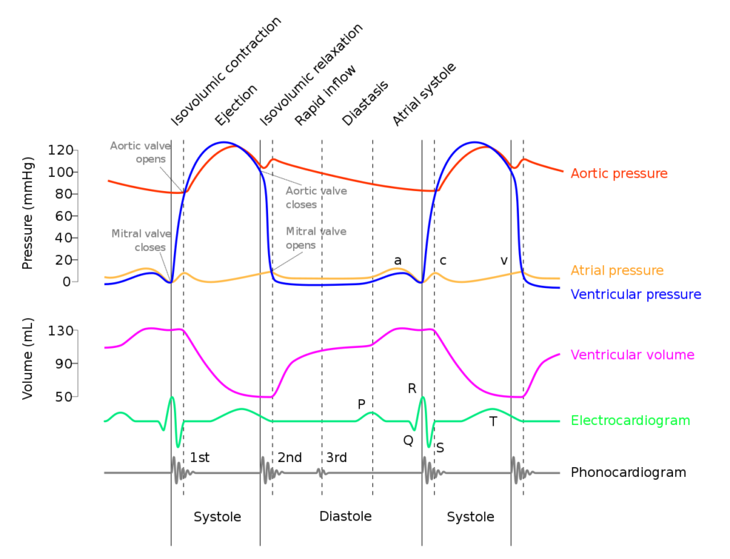
Diastole in the cardiac cycle corresponds with cardiac myocyte relaxation, and includes periods of isovolumetric relaxation and ventricular filling. It lasts approximately 430ms.
Isovolumetric relaxation
- As aortic pressure overcomes ventricular pressure, and the inertia of forward flow of blood out of the LVOT, the aortic valve closes
- The ventricular myocytes relax, while both the mitral (or tricuspid) and aortic (or pulmonary valves) remain closed
- The volume of blood within the ventricle remains constant (LV End-Systolic Volume ~50mls) , however ventricular pressure decreases from 120mmHg to close to 0mmHg (or 20 to 0mmHg in R ventricle
Ventricular filling
- Passive phase
- As ventricular pressure decreases below the atrial pressure, the mitral (or tricuspid) valve opens allowing forward flow of blood from the atria to the ventricles leading to a progressive increase in ventricular volume and pressure.
- The first two-thirds of the ventricular filling is due to passive filling
- Active phase
- 20% of ventricular filling is provided by the atrial kick during the final quarter of ventricular filling resulting in a slight increase in ventricular pressure
- Ventricular pressure overcomes atrial pressure and the mitral (or tricuspid) valves close
Electrochemical events
- The cardiac action potential is tied to contraction via excitation-contraction mechanism.
- Diastole corresponds to phase IV where voltage gated L and T-type Ca channels close, ceasing Ca influx, and ongoing K efflux repolarizes the myocyte.
- ECG
- The T wave corresponds with ventricular repolarization and occurs immediately prior to ventricular diastole.
- The QRS complex corresponds with ventricular depolarization and occurs immediately prior to ventricular systole
- Diastole corresponds with the terminal part of the T wave to immediately after the QRS complex
- Electrical current through cardiac cycles
- During depolarization, current runs from the base of the heart to the apex (corresponding to flow down bundle of His) the just before the end of depolarization, the direction of flow reverses (corresponding to flow down Purkinje fibres)
- During repolarization, the order reverses, from Purkinje system → Bundle of His
Coronary artery perfusion
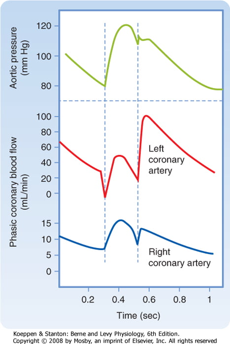
- Due to the high ventricular pressures impeding blood flow during systole in a starling-resistor model, left ventricular perfusion (or right ventricular perfusion during pressure-overloaded states) occurs predominantly during diastole.
- As right ventricular pressures are significantly lower than aortic pressures even during systole, perfusion continues (in a normal right ventricle)
Sakurai 2016
Examiner Comments
2024B 13: 38% of candidates passed this question.
A thorough response to this question provided an accurate definition of ventricular diastole providing detail about duration (either duration at a certain heart rate or the usual ratio with changes due to tachycardia). The second part was best structured by dividing the events into cardiac mechanical and electrical events and circulatory changes. Mechanical events included a description of isovolumetric relaxation, early filling, atrial ejection and isovolumetric contraction, with an explanation of what marks the beginning and end of each of these phases as well as what happens to pressures within the cardiac chambers. Electrical events included the correlation of diastole with ventricular repolarisation and refractory periods as well as the corresponding ECG waves. Finally, an overview of the changes that occur in the coronary, pulmonary and systemic circulations during diastole was expected.
2018A 12: 29% of candidates passed this question.
Many answers lacked structure and contained insufficient information. Better answers defined diastole and described the mechanical events in the 4 phases of diastole. A common error was the ECG events in diastole. The electrical events and coronary blood flow should have been mentioned.
2011A 21: 1 (8%) of candidates passed this question.
One possible way to answer this question is to offer a definition of the diastolic period then to split the events up for description into mechanical events, ECG events and electrical/ionic events.
Few candidates defined the diastolic period, and whilst many talked about opening and closing of valves, there was generally a poor understanding of the sequence of events whereby the left ventricle comes to be filled with blood. The better answers included a description of the ionic events that occurred at the various stages of diastole. Many answers lacked any reference to the ECG events in diastole.
The major weakness in answers was again the failure to include sufficient information to achieve a pass mark. This was probably as a result of the lack of a systematic approach when answering a question of this nature.
Syllabus: C1b, 2d,e and C1c, 2e,f
Recommended sources: Textbook of Medical Physiology, Guyton & Hall, Chp 9 – 11 and Review of Medical Physiology, Ganong, Chp 31
2010B 23
Describe the ionic events associated with a ventricular cardiac action potential (80% of marks).
Outline how the action potential relates with the mechanical events of the cardiac cycle (20 % marks).
CICMWrecks Answer
Normal Values:
- AP Duration 200-250ms
- ARP 200ms
- RRP 50ms
RMP -90
Established and maintained by:
- Na/K-Atpase
- Na/Ca antiporter
- Ca ATPase
Threshold potential -65mv
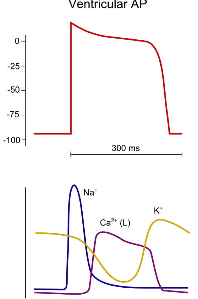
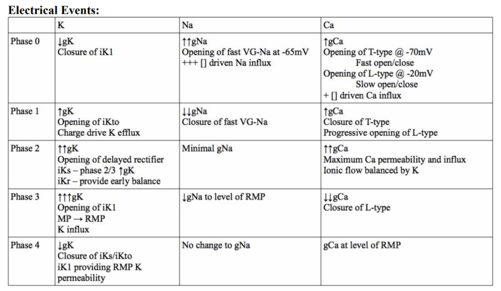
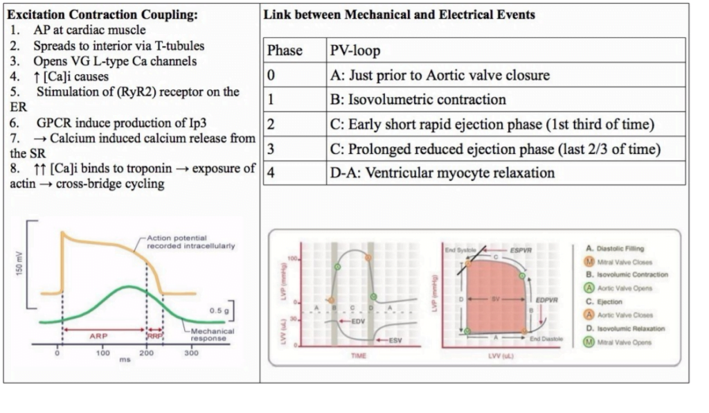
<To be updated>
Gladwin 2016
Examiner Comments
2010B 23: 4 (27%) of candidates passed this question.
To achieve a good pass in this question, candidates needed to outline the ionic events associated with Phase 0 to phase 4 of the ventricular action potential followed by a description of excitation – contraction coupling. The second part of the question was best answered using a ventricular pressure-volume loop and overlaying the phases of the ventricular action potential.
Description of the ionic events associated with the action potential phases was generally well done, but this was as far as many answers went in answering this question. Few candidates included a description of excitation-contraction coupling in there answer and few candidates considered an answer to the second part of the question. The use of illustrations helped answer this question.
Syllabus: C1b
References: Guyton and Hall Textbook of Medical Physiology, Chp 9
2011A 13
Relate the surface electrocardiogram (ECG) to the events of the cardiac cycle (60% of marks). Briefly describe the mechanism of the effects of digoxin, and the mechanism of the effects of amiodarone, on the ECG (40% of marks)
2009A 01
Relate the surface ECG to the events of the cardiac cycle (60% of mark). Describe how the PR, QRS and QT intervals may be prolonged by the action of drugs.
CICMWrecks Answer
Surface ECG to events of Cardiac Cycle
ECG:
- 12 metal electrodes on chest wall
- Detect small (0.5-2mV) changes in voltage produced by the heart
- Transfer these to an oscilloscope output
- P wave: → atrial depolarisation → atrial systole → AV valve opening
- PR interval: conduction from SA node → atrial conducting pathways → AV node (slowest) → bundle of His → LBB and RBB → Purkinje fibres
- QRS: ventricular depolarisation (endocardium → endocardium, L > R) → ventricular systole → closure of AV valves, opening of aortic and pulmonary valves
- Atrial repolarisation is low voltage, and lost within the QRS complex
- QT interval: sustained ventricular depolarisation and muscular contraction
- T wave: ventricular repolarisation (epicardium -> endocardium, L > R) -> ventricular
relaxation -> closure of aortic and pulmonary valves
ELECTRICAL EVENTS
| P0 | Fast Na opening |
| P1 | Transient K efflux |
| P2 | Influx Na & Ca |
| P3 | Efflux K > Influx Na & Ca |
| P4 | Re-establishment of RMP |
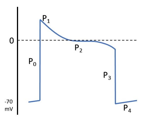
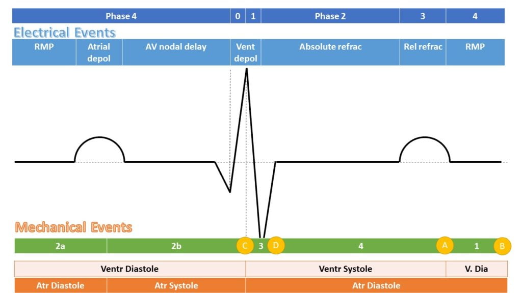
MECHANICAL EFFECTS
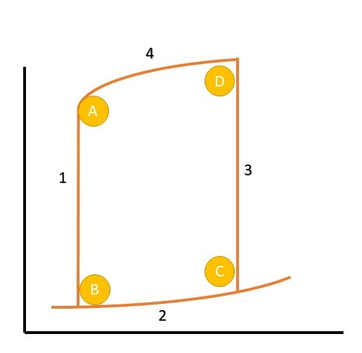
| A | Aortic Valve Closure |
| 1 | Isovolumetric relaxation |
| B | Mitral Valve Opening |
| 2a | Early Diastolic filling |
| 2b | Late Diastolic filling |
| C | Mitral Valve Closure |
| 3 | Isovolumetric Contraction |
| D | Aortic Valve Opening |
| 4 | Ventricular Ejection |
Action of Drugs
| Drug | Interval effects | Mechanism |
|---|---|---|
| VW (Vaughan-Williams) Class 1a | ↑QRS, ↑QT | Fast Na-channel blockade |
| VW Class 1b | ↓QT | |
| VW Class 1c | ↑↑QRS, ↑QT | |
| VW Class 2 | ↑PR | β blockade → negative chronotropy |
| VW Class 3 | ↑QT | K channel blockade → prolongation of RRP |
| VW Class 4 | Can ↑PR | Prolongation of ERP and RRP in pacemaker cells |
| Adenosine | ↑PR | Hyperpolarisation of myocardium via opening of K channels |
| Digoxin | ↑PR, ↓QT | Multiple effects, vagotonic at AVN |
| Amiodarone | ↑PR, ↑QRS, ↑QT, ↑RR (sinus rate) | Multiple |
| Mg | ↓QTc | Membrane stabilising. |
| TCAs | ↑QRS | Quinidine like Na-channel effect (class 1a) |
| SSRIs | ↑QT | Direct potassium blockade and downregulation of potassium → ↑ERP and ↑RRP. |
Digoxin
- ↑ RR interval (sinus rate), ↑PR interval, ↓RR interval
- Slowed AV conduction by increased vagal tone
- ACh → M2 receptors
- ↑KACh (ACh controlled K+ channels) → increased efflux of K+ → hyperpolarised
membrane - ↓If (funny current) → decreased influx of Na+ → decreased slope of phase 4
- ↓ICa(L) (long lasting) and iCa(T) (transient) → decreased efflux of calcium on partial
depolarisation → decreased slope of phase 4
Amiodarone
- ↑PR, ↑QRS, ↑QT, ↑RR (sinus rate)
- Main effect is K+ channel blockade
- Repolarisation is slowed → prolonged QT
- Some INa
- Slower phase 0 upstroke
- Slowed ventricular muscle conduction → prolonged QRS
- Some ICa
- Slower spontaneous depolarisation in SA node → decreased sinus rate
- Slowed conduction in AV node → prolonged PR
- Weakly downregulates adrenoreceptors
- Increased PR, decreased sinus rate
Mooney / Gladwin 2016
Examiner Comments
2011A 13: 8 (66%) of candidates passed this question.
Candidates were expected to provide sufficient detail in answers. Extra marks were awarded for diagrams relating the ECG accurately to pressure events during the cardiac cycle. Time intervals, units of measurement and clear labels were essential for diagrams.
Mechanisms pertaining to ion flux and ion channels needed to be specifically explained. Discussion of mechanisms needed to be accurate and relevant to the effect on the ECG. For example, better answers noted that AV conduction was depressed by Digoxin, predominantly due to an increase in Vagal tone
2009A 01: Pass rate: 50%
The first part of the question is best answered by a labelled and annotated diagram of the ECG with the pressure events of the cardiac cycle. Common errors included mistiming of the ECG with the pressure waveform. The second part of the question could be answered in a tabular format such as:
| Interval | Drug | Mechanism |
|---|---|---|
| PR | Digoxin | Increases refractory period of AV node probably by increased vagal activity |
| QRS | Tricyclic antidepressants | Quinidine like effect, decreasing sodium influx into cells |
| QT | SSRI’s | Malfunction of calcium ion channels |
Syllabus Cib2c, C2c2b
References: Power and Kam 2nd edition p129 – 131, Stoelting and Hillier 4th edition p403, 409, 415

Recent Comments