Syllabus (Fourth Edition, 2023)
Topics
i. Describe the structure and function of the upper airway, lower airway, and alveolus.
ii. Describe the structure of the chest wall and diaphragm and relate this to respiratory mechanics.
iii. Outline the anatomy of the pulmonary and bronchial circulations.
Topics not covered in previous SAQs
ii. Describe the structure of the chest wall and diaphragm and relate this to respiratory mechanics.
iii. Outline the anatomy of the pulmonary and bronchial circulations.
Learning Objectives for the First Part Examination in Intensive Care Medicine
- This will ensure that trainees, tutors, and examiners can work from a common base.
- All examination questions are based around this Syllabus.
- These learning objectives are designed to outline the minimum level of understanding required for each topic.
- The accompanying texts are recommended on the basis that the material contained within them provides sufficient information for trainees to meet the learning objectives.
- Trainees are strongly encouraged to explore the existing and evolving body of knowledge of the Basic Sciences as they apply to Intensive Care Medicine by reading widely.
- For all sections of the syllabus an understanding of normal physiology and physiology at extremes of age, obesity, pregnancy (including foetal) and disease (particularly critical illness) is expected.
- Similarly, for pharmacology, trainees are expected to understand a drug’s pharmacology in these contexts.
- An understanding of potential toxicity and relevant antidotes is also expected.
Definitions
Throughout the document specific wording has been used under the required abilities to indicate the level of knowledge and understanding expected and a glossary of these terms is provided.
Definitions
| Calculate | Work out or estimate using mathematical principles. |
| Classify | Divide into categories; organise, arrange. |
| Compare and contrast | Examine similarities and differences. |
| Define | Give the precise meaning. |
| Describe | Give a detailed account of. |
| Explain | Make plain. |
| Interpret | Explain the meaning or significance. |
| Outline | Provide a summary of the important points. |
| Relate | Show a connection between. |
| Understand | Appreciate the details of; comprehend. |
SAQs
i. Describe the structure and function of the upper airway, lower airway, and alveolus.
2023B 16 – 2020B 14 – 2015A 24
Outline the anatomy of the larynx.
CICMWrecks Answer
Cartilages
- Unpaired
- Thyroid cartilage – Level of C4~5
- Cricoid cartilage
- Epiglottis
- Paired
- Arytenoid
- Cuneiform
- Corniculate
Muscles
- Intrinsic
- Cricothyroid
- Originates in cricoid cartilage and inserts into thyroid
- Tenses vocal cords and elevates voice
- Thyroarytenoid
- Originates in thyroid cartilage and inserts into arytenoid cartilage
- Relaxes vocal cords and depresses voice
- Posterior cricoarytenoid
- Abducts the vocal cords
- Lateral cricoarytenoid
- Adducts the vocal cords
- Oblique and transverse arytenoids
- Adducts the vocal cords
- Cricothyroid
- Extrinsic
- Strap muscles
Innervation
Sensory
- Internal branch of superior laryngeal nerve
Motor
- Cricothyroid – External branch of the superior laryngeal nerve
- All other intrinsic muscles – Recurrent laryngeal nerve
Blood supply
- Superior laryngean artery (branch of external carotid)
- Inferior laryngeal artery (branch of thyrocervical trunk from subclavian artery)
Associations
- The thyroid glands lie inferolateral to the larynx (lateral to the cricoid, the isthmus is inferior to the cricoid)
- The oesophagus, anterior longitudinal ligament, cervical vertebrae lies posterior to the larynx
- The brachiocephalic trunk may arch superiorly close to the cricothyroid membrane in an anatomical variant
Sakurai 2016
Examiner Comments
2023B 16: 44% of candidates passed this question.
This question required candidates to address the following relevant to the anatomy of the larynx – it’s location and extent; relations; structure (paired and unpaired cartilages; major ligaments; intrinsic and extrinsic muscles); nerve supply (sensory and motor); and blood supply (and venous darinage). There were some marks allocated for other correct information relevant to the anatomy of the larynx (e.g. epithelium; differences in age; lymphatic drainage). This was a purely anatomy question so functions of the larynx was not required.
2020B 14: 40% of candidates passed this question.
For this question, candidates were expected to address the location of the larynx, its relations, the cartilages (single and paired), ligaments, muscles (intrinsic and extrinsic), innervation (sensory and muscular) and blood supply (including venous drainage). Marks were apportioned to each section, so whilst some detail was required, breadth of knowledge was also important. Most candidates had a grasp of the gross anatomy, the main relations and at least the innervation provided by the recurrent laryngeal nerve. However, an understanding of the functional anatomy of the cartilages was not always apparent. It should be noted that not every single muscle needed to be named (especially for the extrinsic muscles), but an understanding of the major muscle groups should have been included.
2015A 24: 13 % of candidates passed this question.
It was expected that an answer would include the names of the three single and three paired laryngeal cartilages, intrinsic and extrinsic muscles (names were not required), nerve supply (motor and sensory) and blood supply. Many candidates had good illustrations though a drawing was not essential.
The majority of candidates failed to name the laryngeal cartilages. There was much confusion about whether certain structures were bones or cartilage or even muscle. The relation of the larynx to the thyroid gland was frequently misunderstood.
Many answers focussed on the relations of the larynx but omitted basic information about the larynx itself. No marks were awarded for the contents of the carotid sheath or the course of the recurrent laryngeal nerve both of which were frequently included in answers.
2009B 07
Compare and contrast the anatomy of the upper airway of a term newborn with that of an adult.
CICMWrecks Answer
Miller’s 5 Differences
| Newborn | Adult |
|---|---|
| Disproportionately large tongue that complicates laryngoscopy No dentition | Smaller tongue |
| Larynx is more cephaled (Cricoid at C4) | Larynx more caudal (Cricoid at C6) |
| Epiglottis is shaped differently (U shaped), being short, stubby, omega shaped, and angled over the laryngeal inlet | Epiglottis is longer and stiffer |
| Vocal cords are angled | |
| Larynx is funnel shaped, the narrowest portion occurring at the cricoid cartilage Oedema due to trauma may rapidly cause airway obstruction. |
Other Differences
| Airway | Narrower airways increase the resistance and work of breathing Airway more oedematous due to hormones from mother (oestrogen/progesterone) | Larger airways, less resistance and WOB |
| Nose | Obligate nose breathers Nasal obstruction may significantly impair respiration. | |
| Head | Proportionally enlarged head and occiput Optimal intubating position is neutral rather than ramped. | Head is relatively smaller |
| Neck | Less muscular and more mobile neck Proportionally short neck Favours airway obstruction when flexed. Less effective pharyngeal dilators | Decreased mobility and more likely to have arthritis and pathological issues More effective pharyngeal dilator muscles |
| Trachea | Intrathoracic trachea is also shorter May be only 4cm long, so there is little margin for error in tube placement. | |
| Bronchi | Angles of the bronchi take off is similar making left sided intubation as likely as right | Accidental intubation more likely to be right sided |
| Other differences | Increased physiological dead space due to large head 3.3 ml/kg | Decreased PDS 2 ml/kg |
JC / Gladwin 2020
Examiner Comments
2009B 07: 2 (22%) of candidates passed this question.
Good answers to this question directly compared the anatomical differences using two columns: one for the newborn and the other for the adult airway. They further divided the differences anatomically into mouth/naso and oro pharynx, the glottis (and epiglottis), the larynx, and the trachea (main carina and main bronchi).
Common omissions were failure to mention that neonates do not have dentition, have more compliant tissues and reduced muscle tone, that the neonates’ larynx lies more cephalad and anteriorly, is covered by a large floppy epiglottis and that the main carina also lies more cephalad.
Few candidates were able to list more than 4 differences between the anatomy of the two airways and none mentioned the possibility of disease affecting the neck in the adult.
Syllabus – P2e
Reference: Anatomy for anaesthetists and T K Oh Chp95, Anatomy at a glance.
2019B 05
Describe the anatomical course and relations of the trachea and bronchial tree (to the level of the segmental bronchi).
2016B 24
Outline the tracheal (60% of marks) and left and right main bronchial anatomy (40% of marks) in an adult.
CICMWrecks Answer
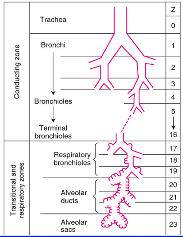
Tracheobronchial Tree
Trachea →
Primary main bronchi →
Lobar bronchi →
segmental bronchi (each supplies one bronchopulmonary segment) →
divide dichotomously→→→→
terminal bronchioles →
respiratory bronchioles →
2-11 alveolar ducts →
alveoli

Trachea
- Midline. 10-12 cm long 15-20mm diameter.
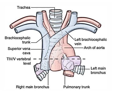
- From Cricoid (C6 level) to bifurcation (T4-T6 level)
- 16-20 C-shaped cartilage rings
- Vertical fibroelastic tissue, and posterior trachealis muscle
- Arterial supply: Upper 2/3 – Inferior thyroid artery, Lower 1/3 Bronchial artery
- Venous drainage: Inferior thyroid vein
- Lymphatics: Drain into deep cervical, pre-tracheal, paratracheal lymph nodes
- Nerve supply: Recurrent laryngeal nerve, Sympathetic fibres from middle cervical ganglion
Bronchi
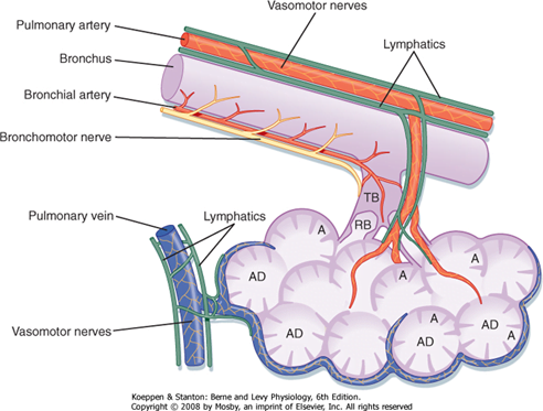
- Supplied by bronchial arteries which run along the bronchi (with vessels from pulmonary circulation)
- Drains into bronchial veins
- Similar innervation as trachea
- A bronchopulmonary segment is a portion of lung supplied by a specific segmental bronchus and arteries.

Right main bronchus
2cm long, I.D. 10-16mm
Shorter, wider, more nearly vertical than left
Branches:
- Right upper lobe
- Apical
- Anterior
- Posterior
- Bronchus intermedius
- Middle lobe
- Medial
- Lateral
- Lower lobe
- Superior
- Medial basal
- Anterior basal
- Lateral basal
- Posterior basal
- Middle lobe
Left main bronchus
4-5cm long, I.D. 8-14mm
Crosses anterior to the esophagus, which it indents.
Branches:
- Left upper lobe
- Apicoposterior
- Anterior
- Lingular
- Superior
- Inferior
- Left lower lobe
- Superior
- Anterior basal
- Lateral basal
- Posterior basal
Lining Epithelium

Trachea → Terminal bronchioles: ciliated pseudostratified columnar epithelium
Respiratory bronchioles → alveolar ducts → alveoli: Non ciliated cuboidal epithelium
Sources: Nunn’s Applied Respiratory Physiology, West’s Respiratory Physiology, Berne and Levy Physiology
JC 2019
Examiner Comments
2019B 05: 14 % of candidates passed this question.
To pass this question, the following were required for each section (trachea and main bronchi): landmarks; basic structural anatomy; and important relations (major vessels; major nerves; major structures). Marks were also allocated for innervation, and blood supply and venous drainage of the
trachea. Most unsuccessful answers did not address a number of these areas. Overall, the answers were better for tracheal anatomy compared to bronchial anatomy. A structured approach to anatomy questions works well and this was again the case (i.e. relations / blood supply / etc.
2016B 24: 24% of candidates passed this question.
Better answers included details of the significant structures related to the cervical and mediastinal trachea and bronchi. The lobar branches and bronchopulmonary segments requiring naming to attract full marks. Many answers lacked sufficient detail or contained inaccuracies regarding vertebral levels and key structural relations. Some candidates discussed the general anatomy of the airway, including the larynx, structure of the airways, blood supply and innervation. This did not attract marks.
2023A 04
Explain the structural features of the alveolus that facilitate its function?
2016A 15
Describe the structure and function of the alveolus.
CICMWrecks Answer
Alveolus
- Hollow cavity found in the lung parenchyma, Ends of the respiratory tree.
- There are ~300 million alveoli that are engulfed by a rich capillary network
- cumulative Surface Area of all alveoli is ~85 m2 with a volume of 4000 mL
- Polyhedral-shaped → 0.33 mm in diameter
- Alveolar walls contain dense mesh of capillaries large enough for a red cell to pass through
- Each alveolar wall facing air is lined by Type I cells (one-cell-thick) → parts of alveolar
wall consists of interstitial space that contain pulmonary capillaries - Diffusion barrier that alveolar gas has to travel to pulmonary blood is short (0.3 um only!)
→ consists of (i) Type I cell (epithelium), (ii) Basement membrane of type I cell, (iii)
Basement membrane of pulmonary capillary endothelium, and (iv) Pulmonary capillary
endothelium - In some alveolar walls, there are pores between alveoli called Pores of Kohn
- Alveoli are inherently unstable (Ie. collapse) given their small size and surface tension of
their liquid-lining → this is offset by surfactant produced by Type II cells (round cells
with large nuclei and cytoplasmic granules containing surfactant)
Alveolar cells
Type 1 Pneumocytes
- 90% of alveolar surface area
- Thin walled, Involved in gas exchange
- Unable to replicate, Susceptible to toxic insults,
Type II pneumocytes
- 10% of alveolar surface area
- Secrete surfactant
- Proliferate and differentiate into type I and type II cells
Alveolar Macrophages
- Present in alveolar septae, lung interstitium
- Phagocytosis of foreign particles
JC / Bianca 2019
Examiner Comments
2023A 04: 14% of candidates passed this question.
The question asked for an explanation of the structural features of the alveolus that facilitate its function. Candidates who scored well integrated the specific anatomical and structural elements of the alveolus with the multiple functions of the alveolus, including the relevant explanation regarding mechanisms. No marks were given for simply listing the structural features of an alveolus without an explanation on how this facilitates function, nor for listing functional requirements of an alveolus without explaining whether or how they are met by its structure. For the same reason, this was one question where simply listing equations with no discussion as to how these relate to the structure and function of the alveoli garnered no marks. Equations were not required for full marks but may be an efficient way to represent physical relationships that are hard to write in a few words. Common omissions included a description and the role of collagen and elastin fibres, capillary structure, the filtration function of the membrane, lymphatic drainage, recruitment and distensibility and metabolic functions. Candidates are encouraged to practice model answer templates for these integrative questions in the months leading up to the exam.
2016A 15: 52% of candidates passed this question.
Better answers related structure to function. Many answers lacked key anatomical features (for example pores of Kohn, basement membrane, interconnecting walls / alveolar interdependence etc.). There was little understanding of the role and origin of the basement membrane of the alveolus. Some candidates went into detailed discussions of Work of Breathing, respiratory mechanics and the renin-angiotensin system which were not asked for.
Answers not reaching a pass mark generally suffered from lacking detail and suggested only a superficial understanding of the area.
ii. Describe the structure of the chest wall and diaphragm and relate this to respiratory mechanics.
2015B 21
Outline the anatomy of the diaphragm (70% of marks).
Describe the function of the diaphragm in respiration (30% of marks).
2011A 02
Outline the principal anatomical features of the diaphragm that are important to its function.
CICMWrecks Answer
Anatomy of the Diaphragm
- Insertion
- Anterior
- Xiphoid process
- Costal cartilage of ribs 6~12
- Posterior
- Lateral arcuate ligament (overlies quadratus lumborum)
- Medial arcuate ligament (overlies psoas major)
- Right and left crux (blends with anterior longitudinal ligament from vertebral bodies L1~3 on right, L1~2 on left)
- Median arcuate ligament (between right and left crux, overlying aorta)
- Central tendon – blends with pericardium superiorly and fibrous capsule of liver inferiorly
- Anterior
- Openings
- Caval orifice (T8) – Within central tendon – Passage of the IVC
- Oesophageal hiatus (T10) – Within sling of right crura – Passage of oesophagus and vagus nerve
- Aortic hiatus (T12) – Posterior to median arcuate ligament between right and left crura – Passage of aorta, azygous vein and thoracic duct
- Innervation
- Motor – Phrenic nerve (C3~5)
- Sensory – Phrenic and inferior intercostal nerves
- Blood supply
- Branches of the lower intercostal arteries, superior phrenic arteries
- Inferior phrenic artery (branch of aorta) from inferior surface
Function of the Diaphragm
- On inspiration
- Diaphragm contracts
- Dome of diaphragm flattens and descends
- Pushes lower thoracic ribs outwards
- Increases AP diameter and superior-inferior distance of thorax → increased thoracic volume → Negative intrathoracic pressure → inspiration of air
- Caval orifice dilates → increases venous return (Liver compressed squeezing hepatic blood reservoir, increased intraabdominal pressure due to descending diaphragm)
- Aortic hiatus constricts → prevents arterial blood regurgitation
- Oesophageal hiatus constricts → aids lower oesophageal sphincter and prevents regurgitation of gastric contents
- Diaphragm contracts
- On expiration
- Diaphragm relaxes
- Elastic recoil of lung causes deflation and expiration
- Diaphragm relaxes
Sakurai 2016
Examiner Comments
2015B 21: 27% of candidates passed this question.
The diaphragm is the principal muscle of respiration. Important and unique features of its anatomy include a central tendon that blends with the pericardium above and the fibrous capsule of the liver below, arcuate ligaments and crura that are important points of muscle insertion. There are also three major and three minor openings that allow passage of structures between the thoracic and abdominal cavities. Candidates who had studied anatomy of the diaphragm were clearly distinguishable from those who had not.
Candidates who followed a traditional template for anatomy answers scored better, providing answers that covered the breath of the topic.
2011A 02: 3 (25%) of candidates passed this question.
Most candidates had a basic knowledge of diaphragmatic function however were uncertain of anatomy and rarely related the two. Candidates were expected to describe the attachments of the diaphragm, openings, nerve supply, actions, including it’s role upon the oesophageal sphincter
Syllabus: B1b 2c
Recommended sources: Anatomy for Anaesthetists, Ellis and Feldman, pages 317 – 323

Recent Comments