Past Papers · SAQ
Tracheal Anatomy & Percutaneous Tracheostomy
2026A Q08
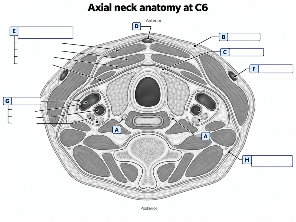
Exam questiona) On the diagram below, identify the anatomical structures labelled A-H (40% of marks).
b) Describe the structural anatomy of the trachea, excluding the anatomical relations (45% of marks).
c) Outline the ideal location for the insertion of percutaneous tracheostomy tube (15% of marks).
CICMWrecks answer
Master answer
a) Diagram labels
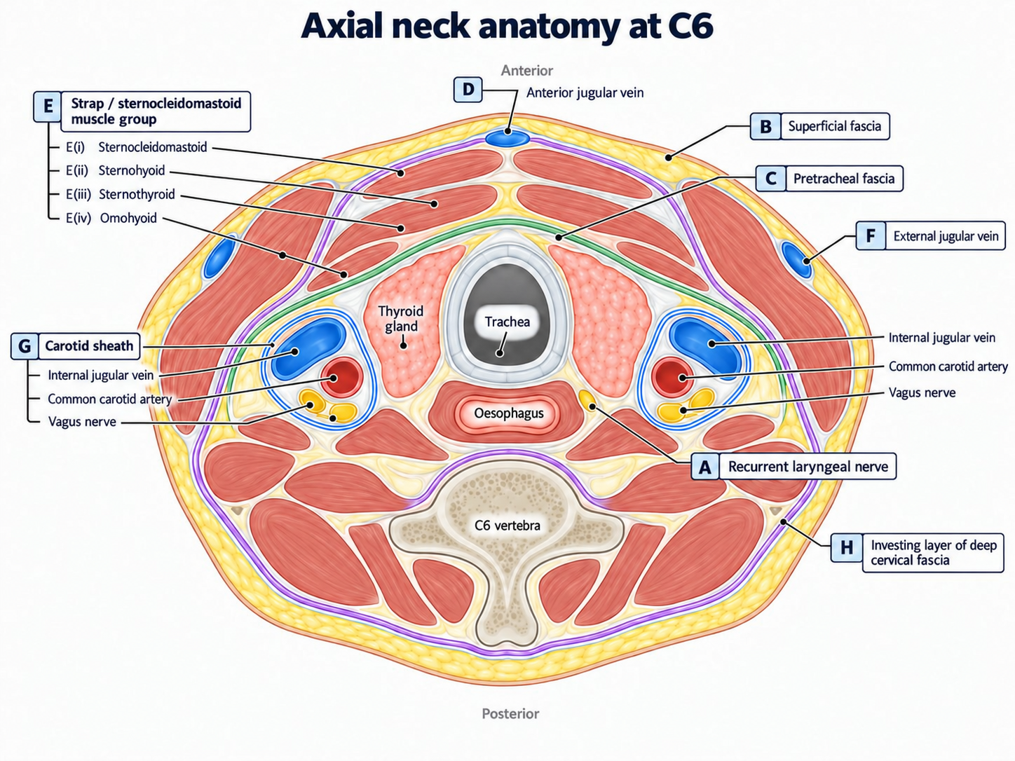
| Label | Structure |
|---|---|
| A | Recurrent laryngeal nerve |
| B | Superficial fascia |
| C | Pretracheal fascia |
| D | Anterior jugular vein |
| E(i) | Sternocleidomastoid |
| E(ii) | Sternohyoid |
| E(iii) | Sternothyroid |
| E(iv) | Omohyoid |
| F | External jugular vein |
| G | Carotid sheath — internal jugular vein, common carotid artery and vagus nerve |
| H | Investing layer of deep cervical fascia |
b) Structural anatomy of the trachea
| Feature | Description |
|---|---|
| Origin | Continuation of the larynx at the lower border of the cricoid cartilage, approximately C6 |
| Termination | Bifurcates at the carina, approximately at the sternal angle / T4–T5 |
| Length | Approximately 10–12 cm in an adult; cervical and thoracic portions |
| Diameter | Approximately 2–2.5 cm in an adult |
| Framework | Approximately 16–20 C-shaped hyaline cartilage rings, incomplete posteriorly |
| Between rings | Adjacent cartilages are joined by fibroelastic annular ligaments |
| Posterior wall | Membranous wall; posterior ends of the cartilage rings are bridged by trachealis smooth muscle and fibroelastic tissue |
| Mucosa | Pseudostratified ciliated columnar respiratory epithelium with goblet cells |
| Submucosa | Connective tissue containing vessels, nerves and seromucous glands |
| Outer layer | Adventitia anchors the trachea to surrounding connective tissue |
| Cartilage function | Maintains luminal patency while allowing flexibility |
| Trachealis function | Changes tracheal calibre; contraction can narrow the lumen and increase expiratory airflow during cough |
Blood supply and innervation
- Arterial supply: cervical trachea mainly via branches of the inferior thyroid arteries; thoracic trachea via bronchial arteries.
- Venous drainage: inferior thyroid venous plexus and bronchial veins.
- Parasympathetic: vagal fibres, including recurrent laryngeal branches.
- Sympathetic: fibres from cervical and upper thoracic sympathetic ganglia.
c) Ideal site for percutaneous tracheostomy
- Midline anterior approach below the cricoid cartilage.
- Insert through an intercartilaginous space within the first three tracheal rings.
- In routine practice, the 2nd–3rd tracheal ring interspace is commonly targeted.
- Avoid an excessively high puncture close to the cricoid because of subglottic/cartilage injury and stenosis.
- Avoid an excessively low puncture because of increasing proximity to the brachiocephalic vessels and mediastinum.
- Remain strictly midline to reduce vascular and recurrent laryngeal nerve injury.
Quick reference
Summary
| Area | Exam anchor |
|---|---|
| Extent | C6 → carina at T4–T5 |
| Framework | 16–20 C-shaped hyaline cartilage rings |
| Posterior wall | Trachealis muscle |
| Lining | Pseudostratified ciliated columnar respiratory epithelium |
| Cervical blood supply | Inferior thyroid arteries |
| Percutaneous site | Midline; usually 2nd–3rd ring interspace |
Past papers
Exam appearances
| Exam | Exact exam wording | Candidate success |
|---|---|---|
| 2026A Q08 | a) On the diagram below, identify the anatomical structures labelled A-H (40% of marks). b) Describe the structural anatomy of the trachea, excluding the anatomical relations (45% of marks). c) Outline the ideal location for the insertion of percutaneous tracheostomy tube (15% of marks). | 38.6% |